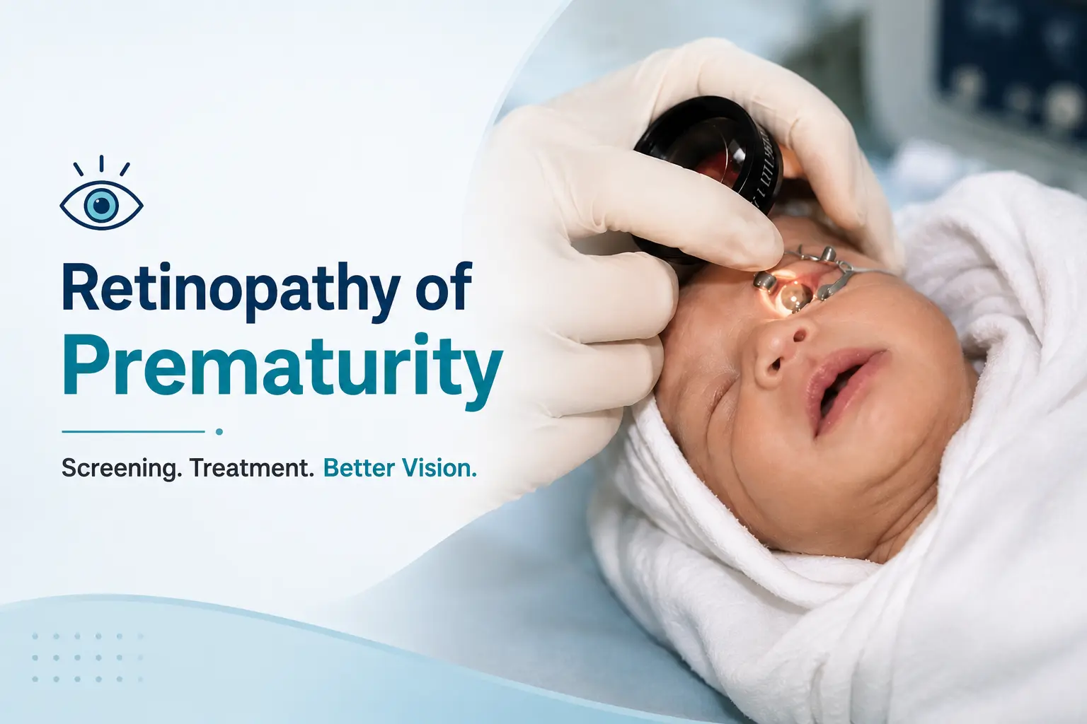
Retinopathy of Prematurity: Screening and Treatment Guide
Retinopathy of Prematurity (ROP) is a fibrovascular proliferative disorder which affects the developing peripheral retinal vasculature of premature infants. Improved neonatal care and better neonatal survival rate has led to the diagnosis of increasing number of cases of ROP. Identifying and screening of at-risk premature infants by an experienced ophthalmologist is the most important strategy in the management of ROP. ROP is a preventable cause of blindness and remains one of the leading causes of visual loss in children. Studies support that both prematurity and supplemental oxygen therapy seem to be important factors in the development of ROP. Screening pictures of retina taken by Retcam can be sent via telemedicine to higher centers and a diagnosis made. The mainstay of treatment has been laser photocoagulation to the avascular retina, although anti VEGF injections have shown promising results.
With the rising rate of prematurity and improving survival, the need for ongoing ROP screening & treatment and long-term follow up is greater than ever.
INCIDENCE
In India, incidence of ROP varies from 38 to 51.9 % among low birth weight babies. Approximately 2 million babies out of 26 million live births annually are born with birthweight <2000 gms and are at risk of developing ROP. According to WHO, India has the highest number of preterm births in the world
Retinopathy of Prematurity Risk Factors
- Low Gestational age
- Low Birth weight
- Number of days oxygen administered
WHEN TO SCREEN
Initial eye examination should be done at 31 weeks PMA or 4 weeks of chronological age whichever is earlier. The first retinal examination should be done in the first month of life.
HOW TO SCREEN
Screening is done in a temperature-controlled room or a nursery in the presence of a neonatologist. Such babies are susceptible to decrease in heart rate and fall in oxygen saturation. A monitor should be there to monitor the heart rate and oxygen saturation throughout the course of examination.
INSTRUMENTATION
1.Retcam-It is a digital camera for screening, provides instant and accurate documentation and provides state of the art wide field pediatric retinal imaging (130 degrees)
2.If a retcam is not available an indirect ophthalmoscope can be used.
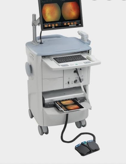
Figure 1a- Retcam
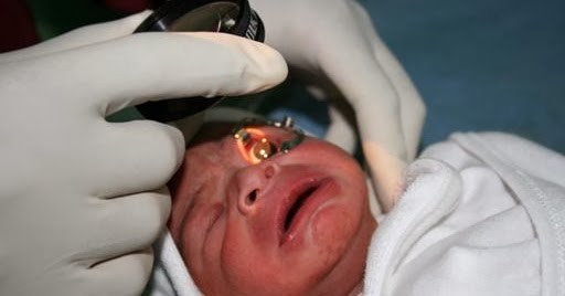
Figure 1b- Examination with indirect ophthalmoscope
HOW TO TREAT
Different modalities of treatment are-
1.Cryotherapy-This modality of treatment is rarely used these days.
2.Laser photocoagulation-This the most widely used modality of treatment. Peripheral laser is done to stop new blood vessel formation. If done adequately and at the right time, the disease regresses and does not come back. The success rate has been quoted to be over 90 percent. It is usually done under topical anesthesia in the presence of an anesthetist, in NICU, with the monitors in place.
3.Anti -VEGF Therapy– VEGF plays a significant role in the disease, and anti -VEGFs play an important role in treatment of the disease. It is easy to use and has a rapid response, sometimes the result is evident within a day of injection whereas it usually takes a week to see the result of laser therapy. Another advantage of anti -VEGF therapy is that it promotes near normal vessel formation and hence preserves peripheral field of vision.
4.Surgery-Indications for surgery are partial and total retinal detachment. Lens sparing vitrectomy has shown good results in stage 4.
SEQUELAE
ROP babies have a strong association with development of myopia. About 65% develop myopia by 9 months of age. These patients also have a high incidence of amblyopia, strabismus and nystagmus. Parents of these babies should be explained the possibility of development of all these sequelae in their childhood years, hence the importance of regular eye checkups.
EXAMINATION SCHEDULE
Following schedule should be followed for babies who do not need ablative treatment:
One week or less follow up
- Stage 1 or 2, zone 1 ROP
- Stage 3, zone 2 ROP
One to two week follow up-
- Immature vascularization zone 1—No ROP
- Stage 2, zone 2 ROP
- Regressing ROP, zone 1
Every two week follow up
- Stage 1, zone 2 ROP
- Regressing ROP zone 2
Every two-three week follow up-
- Immature vascularization zone 2-No ROP
- Stage 1or 2 zone 3 ROP
- Regressing ROP zone 3
Follow up examinations are done till complete retinal vascularization.